| While the Stafford is a relatively disease free breed, there are some serious conditions that plague the breed and all breeders should be testing for. |
|
L-2 - HGA L-2-HGA (L-2-hydroxyglutaric aciduria) in Staffordshire Bull Terriers is a neurometabolic disorder characterised by elevated levels of L-2-hydroxyglutaric acid in urine, plasma and cerebrospinal fluid. L-2-HGA affects the central nervous system, with clinical signs usually apparent between 6 months and one year (although they can appear later). Symptoms include epileptic seizures, "wobbly" gait, tremors, muscle stiffness as a result of exercise or excitement and altered behaviour. The mutation, or change to the structure of the gene, probably occurred spontaneously in a single dog but once in the population has been inherited from generation to generation like any other gene. The disorder shows an autosomal recessive mode of inheritance: two copies of the defective gene (one inherited from each parent) must be present for a dog to be affected by the disease. Individuals with one copy of the defective gene and one copy of the normal gene - called carriers - show no symptoms but can pass the defective gene onto their offspring. When two apparently healthy carriers are crossed, 25% (on average) of the offspring will be affected by the disease, 25% will be clear and the remaining 50% will themselves be carriers The mutation responsible for the disease has recently been identified at the Animal Health Trust. Using the information from this research, we have developed a DNA test for the disease. This test not only diagnoses dogs affected with this disease but can also detect those dogs which are carriers, displaying no symptoms of the disease but able to produce affected pups. Carriers could not be detected by the tests previously available, which involved either a blood or urine test detecting elevated levels of L-2-hydroxyglutarate or magnetic resonance imaging. Under most circumstances, there will be a much greater number of carriers than affected animals in a population. It is important to eliminate such carriers from a breeding population since they represent a hidden reservoir of the disease that can produce affected dogs at any time. The test is available now. Breeders will be sent results identifying their dog as belonging to one of three categories: CLEAR: the dog has 2 copies of the normal gene and will neither develop L-2-HGA, nor pass a copy of the L-2-HGA gene to any of its offspring. CARRIER: the dog has one copy of the normal gene and one copy of the mutant gene that causes L-2-HGA. It will not develop L-2-HGA but will pass on the L-2-HGA gene to 50% (on average) of its offspring. AFFECTED: the dog has two copies of the L-2-HGA mutation and is affected with L-2-HGA. It will develop L-2-HGA at some stage during its lifetime, assuming it lives to an appropriate age. Carriers can still be bred to clear dogs. On average, 50% of such a litter will be clear and 50% carriers; there can be no affected produced from such a mating. Pups which will be used for breeding can themselves be DNA tested to determine whether they are clear or carrier. |
|
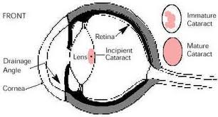
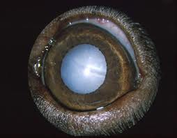
Hereditary Cataract Hereditary Cataract in Staffordshire Bull Terriers has been recognised as an inherited condition since the late 1970’s. Affected dogs develop cataracts in both eyes at an early age. The condition is not congenital, so the lenses are normal at birth, but cataracts appear at a few weeks to months in age, progressing to total cataract (and resulting blindness) by 2 to 3 years of age. The mutation, or change to the structure of the gene, probably occurred spontaneously in a single dog but once in the population has been inherited from generation to generation like any other gene. The disorder shows an autosomal recessive mode of inheritance: two copies of the defective gene (one inherited from each parent) must be present for a dog to be affected by the disease. Individuals with one copy of the defective gene and one copy of the normal gene - called carriers - show no symptoms but can pass the defective gene onto their offspring. When two apparently healthy carriers are crossed, 25% (on average) of the offspring will be affected by the disease, 25% will be clear and the remaining 50% will themselves be carriers. The mutation responsible for the disease has recently been identified at the Animal Health Trust. Using the information from this research, we have developed a DNA test for the disease. This test not only diagnoses dogs affected with the disease but can also detect those dogs which are carriers, displaying no symptoms of the disease but able to produce affected pups. Under most circumstances, there will be a much greater number of carriers than affected animals in a population. It is important to eliminate such carriers from a breeding population since they represent a hidden reservoir of the disease that can produce affected dogs at any time.
The test is available now and information on submitting samples is given below. Breeders will be sent results identifying their dog as belonging to one of three categories: CLEAR: the dog has 2 copies of the normal gene and will neither develop Hereditary Cataract, nor pass a copy of the Hereditary Cataract gene to any of its offspring. Carriers can still be bred to clear dogs. On average, 50% of such a litter will be clear and 50% carriers; there can be no affected produced from such a mating. Pups which will be used for breeding can themselves be DNA tested to determine whether they are clear or carrier. |
|
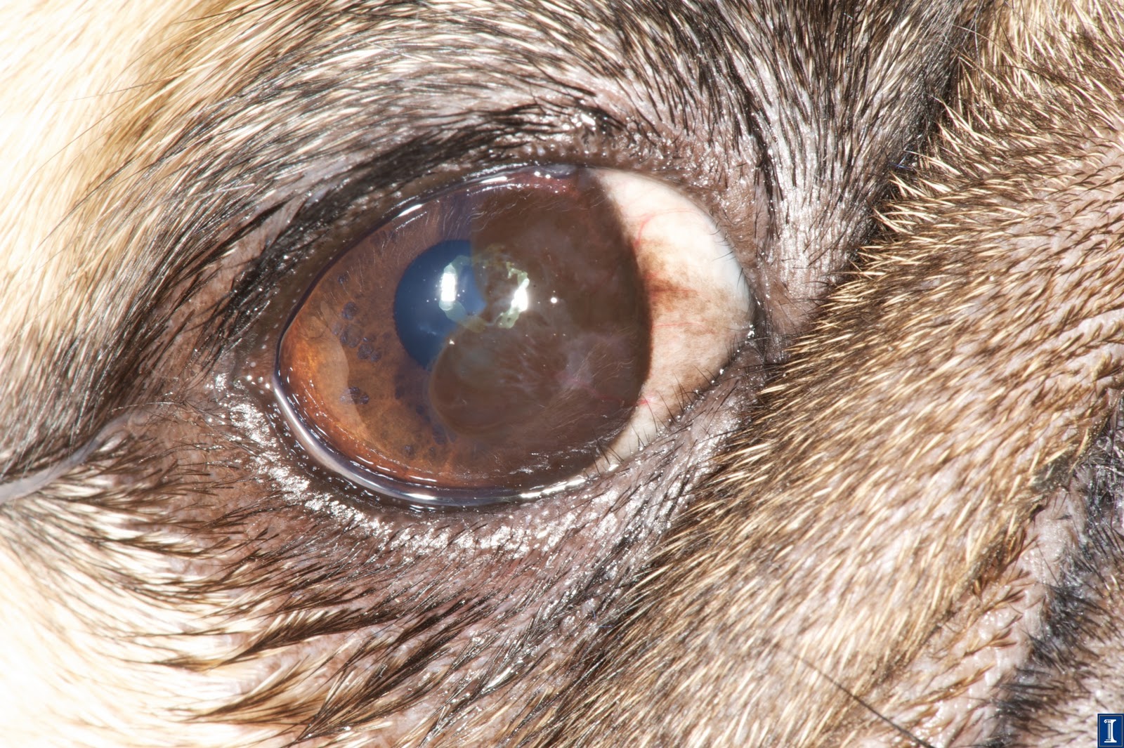
Progressive Retinal Atrophy Progressive retinal atrophy, or PRA as it is frequently termed, is a long recognized, hereditary, blinding disorder. It is inherited as a simple autosomal recessive in most breeds. PRA is a disease of the retina. This tissue, located inside the back of the eye, contains specialized cells called photoreceptors that absorb the light focused on them by the eye’s lens, and converts that light, through a series of chemical reactions into electrical nerve signals. The nerve signals from the retina are passed by the optic nerve to the brain where they are perceived as vision. The retinal photoreceptors are specialized into rods, for vision in dim light (night vision), and cones for vision in bright light (day and colour vision). PRA usually affects the rods initially, and then cones in later stages of the disease. In human families, the diseases equivalent to PRA (in dogs) are termed retinitis pigmentosa. In all canine breeds PRA has certain common features. Early in the disease, affected dogs are night blind, lacking the ability to adjust their vision to dim light; later their daytime vision also fails. As their vision deteriorates, affected dogs will adapt to their handicap if their environment remains constant, and they are not faced with situations requiring excellent vision. At the same time the pupils of their eyes become increasingly dilated, in a vain attempt to gather lighter, causing a noticeable "shine" to their eyes; and the lens of their eyes may become cloudy, or opaque, resulting in a cataract. Diagnosis of PRA is normally made by ophthalmoscopic examination. This is undertaken using an instrument called an indirect ophthalmoscope and requires dilatation of the dog’s pupil by application of eyedrops. Broadly speaking, all forms of PRA have the same sequence of ophthalmoscopic changes: increased reflectivity (shininess) of the fundus (the inside of the back of the eye, overlain by the retina); reduction in the diameter and branching pattern of the retina’s blood vessels; and shrinking of the optic nerve head (the nerve connecting the retina to the brain). These changes occur in all forms of PRA, but at different times in the different breed-specific forms. Usually by the time the affected dog has these changes there is already significant evidence of loss of vision. |
|
Distichiasis Eyelids of dogs can grow abnormal hairs. These hairs grow from the oil glands (Meibomian glands) of the lids and are called distichia if the hair protrudes from the oil gland opening onto the edge of the eyelid. Distichia are often irritating, especially if the hairs are long and stiff. Ectopic cilia are also hairs growing from oil glands on the eyelid, but the hair protrudes from the inner surface of the eyelid and is very painful, often causing corneal ulcers. Dogs with distichiasis may or may not show signs of discomfort, ranging from slight intermittent squinting and/or rubbing of the eyes, to severe squinting and discomfort. Dogs with ectopic cilia are always uncomfortable. Most dogs with ectopic cilia are young adult dogs or older puppies. Both conditions are common in Shih Tzus. Many other breeds have problems with distichia. At Animal Eye Care, both conditions are treated surgically under general anesthesia, with a procedure called cryoepilation. With this procedure, the abnormal hair follicles are frozen using a liquid nitrogen probe, and the hairs then removed. After surgery, the eyelids are swollen for 4-5 days, and the eyelid margins will depigment and turn pink. Usually, the lid margins will repigment within 4 months. It is important to understand that new abnormal hairs can grow from new sites after surgery, but this is uncommon in dogs older than 3 years old (unless the dog is a Shih Tzu). With cryoepilation, 85-90% of the treated hair follicles will not regrow. Repeat surgical treatment is rarely required, unless the animal is a puppy (and grows new hairs in new sites) or a Shih Tzu.
|
|
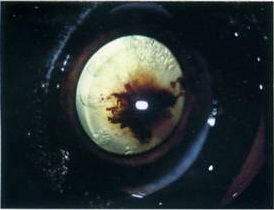
Persistent Hyperplastic Primary Vitreous This is a congenital condition (present from birth) in which there is a developmental defect in the normal regression of some of the intraocular structures of the eye. PHPV can range from being very mild to severe abnormalities which may lead to blindness. The presence of mild abnormalities is usually seen as small brown pigmented dots on the posterior lens capsule. Previously the literature indicated that this was always observed as a bilateral phenomenon but recently it has been stated that affected dogs may show unilateral involvement, although this is less common. The present knowledge of the mode of inheritance of this disease is thought to be an autosomal irregular dominant with variable expression. Due to PHPV seldom resulting in secondary cataracts in the Stafford, those that are mildly afflicted will seldom show any form of visual impairment during their lives. Even those that are more severely afflicted, may be capable of adapting by using peripheral to compensate.
Stafford breeders should therefore not assume that the problem is absent simply because they have not encountered blatant signs of visual impairment, instead discerning breeders should ensure that all their Staffords are tested through the National Eye Scheme. |
|
Entropion Entropion is a genetic condition in which a portion of the eyelid is inverted or folded inward. This can cause an eyelash or hair to irritate and scratch the surface of the eye, leading to corneal ulceration or perforation. It can also cause dark-coloured scar tissue to build up over the wound (pigmentary keratitis). These factors may cause a decrease or loss of vision.
Entropion is common in dogs and is seen in a wide variety of breeds, including short-nosed breeds, giant breeds, and sporting breeds. Entropion is almost always diagnosed around the time a puppy reaches its first birthday. |
|
Ectropion Primarily an inherited condition, in which the lower eyelid droops away from the eyeball to expose the third eyelid and the conjunctiva. Exposure of the delicate mucous membrane causes conjunctivitis.
Correction is possible by complicated surgery in which the eyelid is lifted and shortened. Occasionally further surgery may be necessary to change completely the shape of the eyelids. |

JSN Tendo template designed by JoomlaShine.com